The microorganisms that live in hypersaline lakes are considered to be extremophiles. An extremophile is a living creature, usually microbial,
that thrives under conditions that humans cannot tolerate. Hypersaline lakes have large amounts of salt dissolved in them at higher concentrations
than seawater. These lakes can also range in pH values from extremely acidic to extremely basic. So the microorganisms in these lakes are able to
thrive under very salty conditions and acidic to basic conditions.
Photo taken by Melanie Mormile
Many hypersaline environments have been previously studied by growing microbial cultures and analyzing DNA. These environments have included
areas such as the Dead Sea in Israel, Mono Lake in California, Soap Lake in Washington State, and Lake Magadi, Kenya. However, most of these
lakes are hypersaline environments with neutral to basic pH.
There have been very few reports on microbial studies on the hypersaline lakes
of Victoria and Western Australia and those that have been done have mostly focused on microorganisms from lakes that have neutral to basic pH
values. The lakes in Australia present a great opportunity to study extremophiles that can thrive under both very salty and acidic conditions.
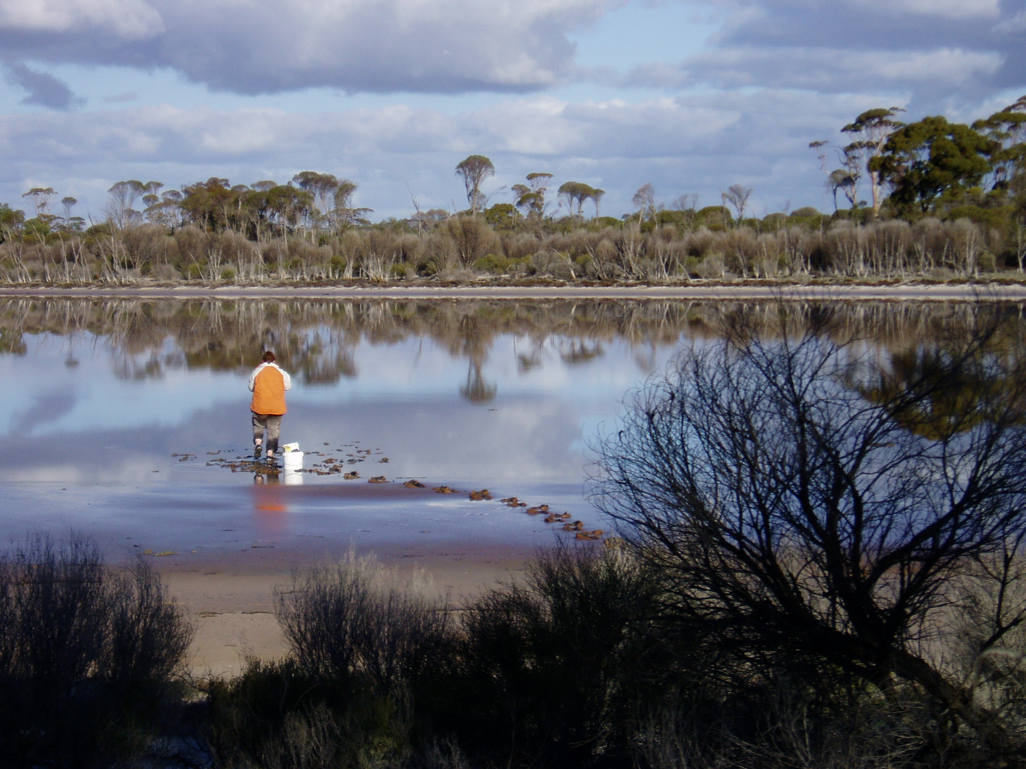
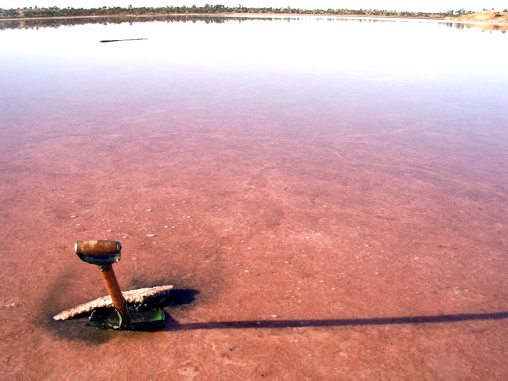
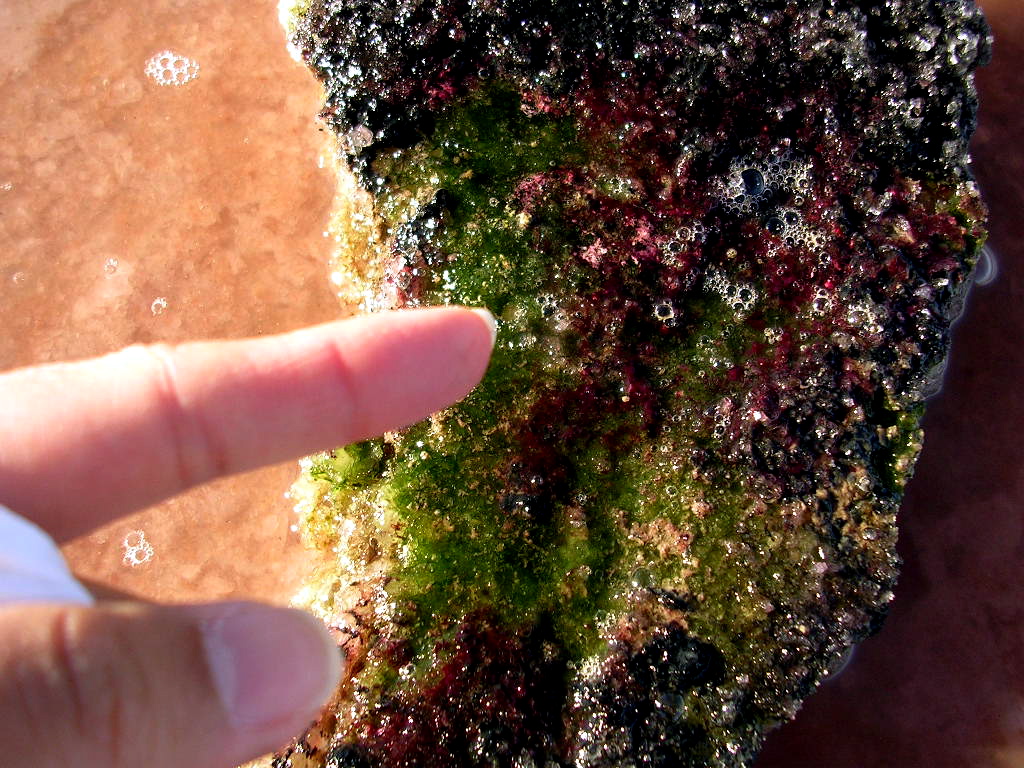
There are many indicators that microorganisms are present when you look around a hypersaline lake. One indicator is color. Many halophiles, salt-loving
microorganisms, have a bright pink to orange color. This causes many of these lakes to look pink. In the photo below, the bottom of this lake has a salt crust.
Microorganisms are trapped in the salt causing it to become pink.
Photo taken by Bo-young Hong
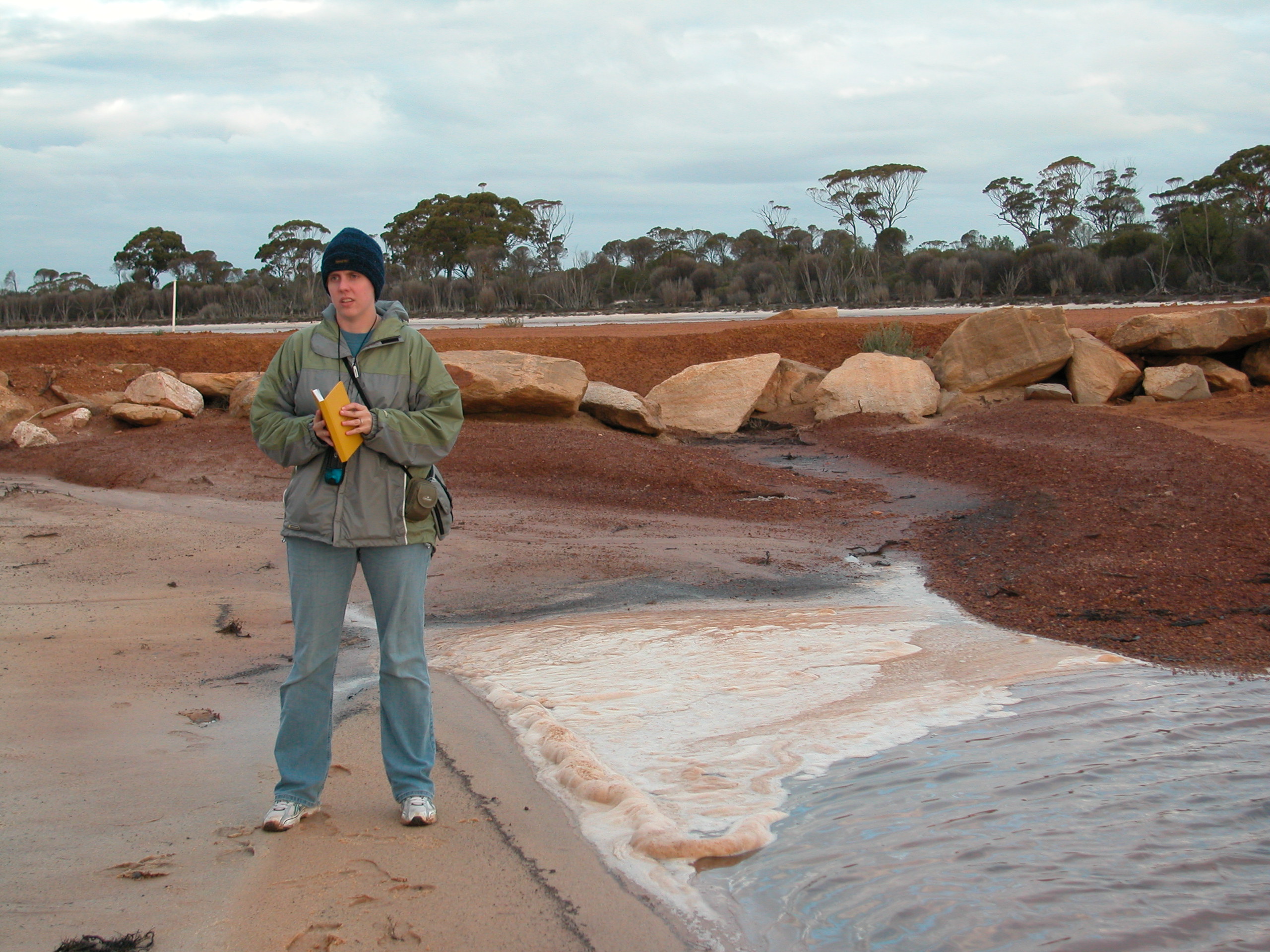
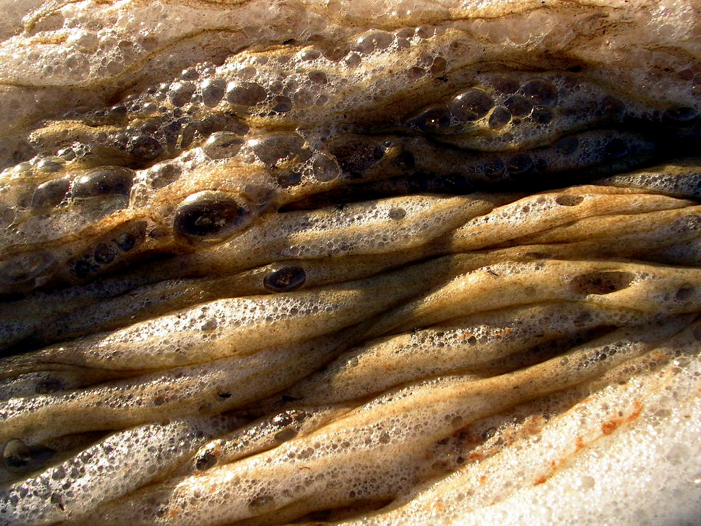
When walking along the shore of a hypersaline lake, you can also notice foam and bubbles. These show that there is microbial activity going on.
Photo taken by Francisca Oboh-Ikuenobe
The slime is made up of the microorganisms and their products. Many microbes produce slime to protect themselves.
Photo taken by Bo-young Hong
The bubbles show that the microbes are producing gas. If they are photosynthetic, they are probably producing oxygen.
Photos taken by Melanie Mormile
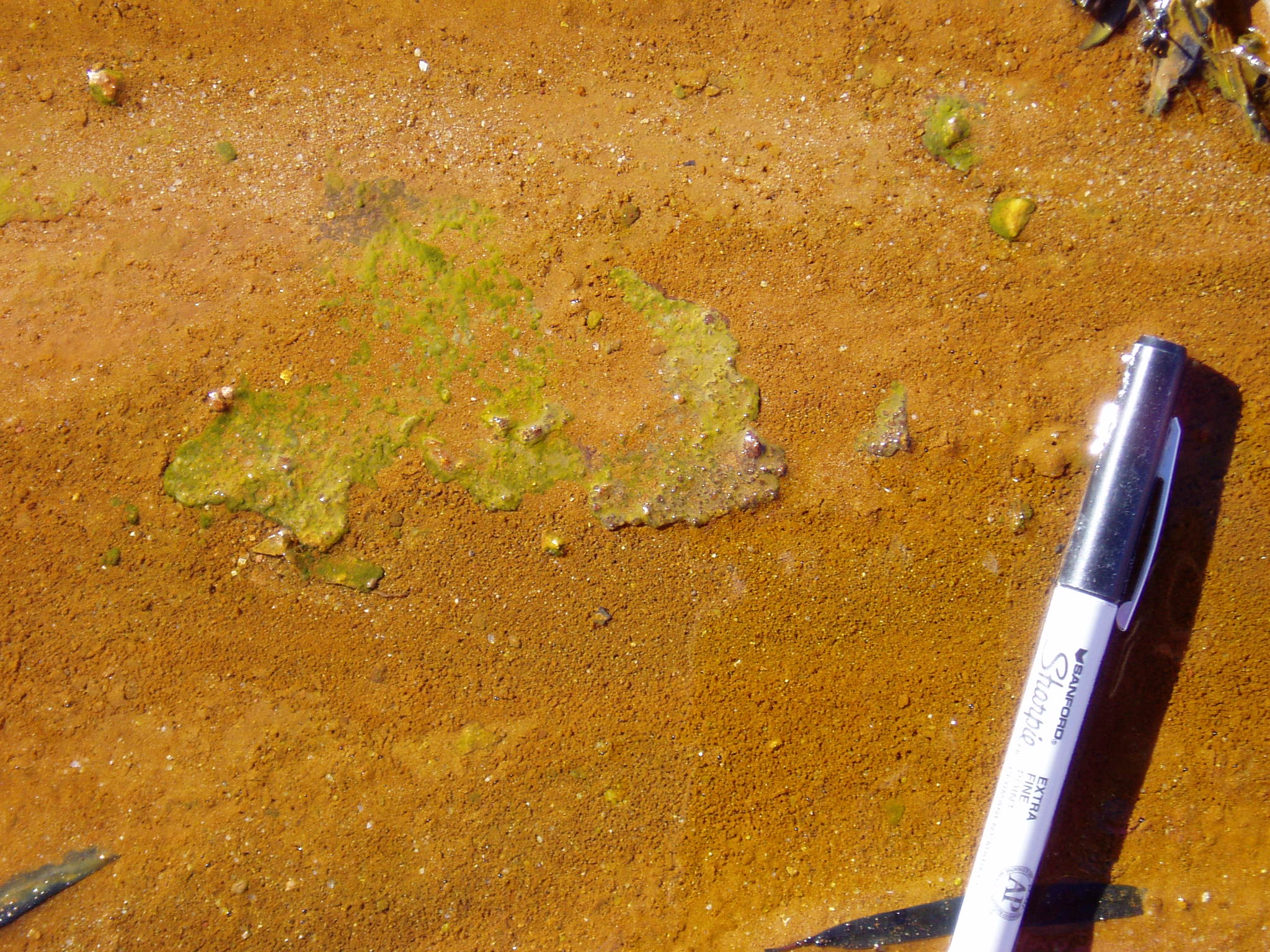
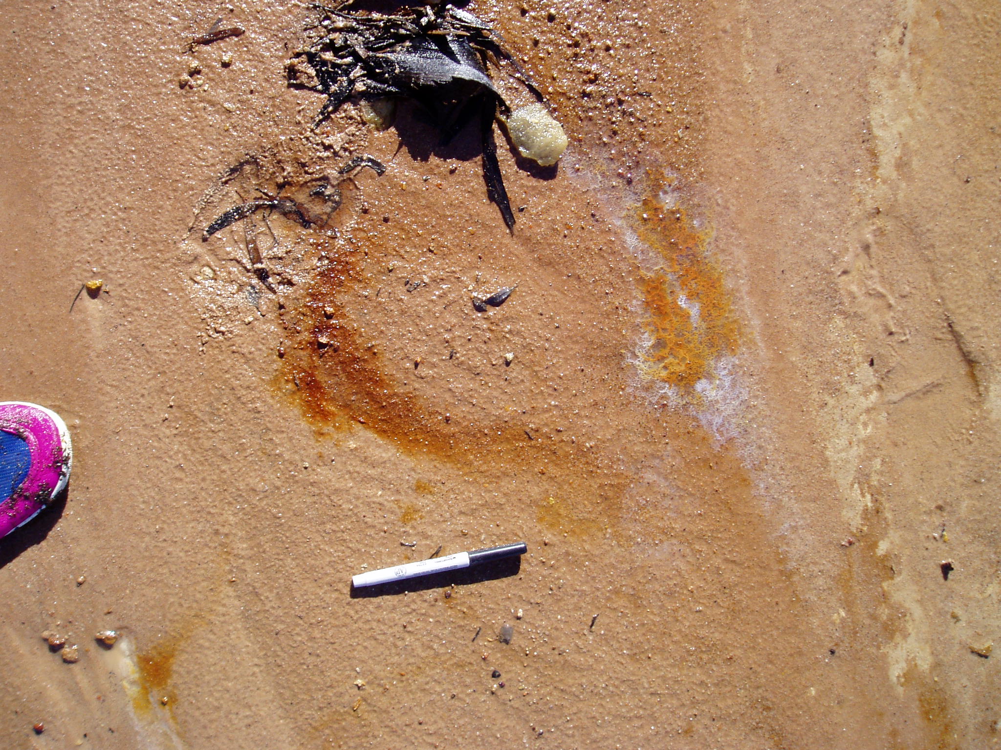
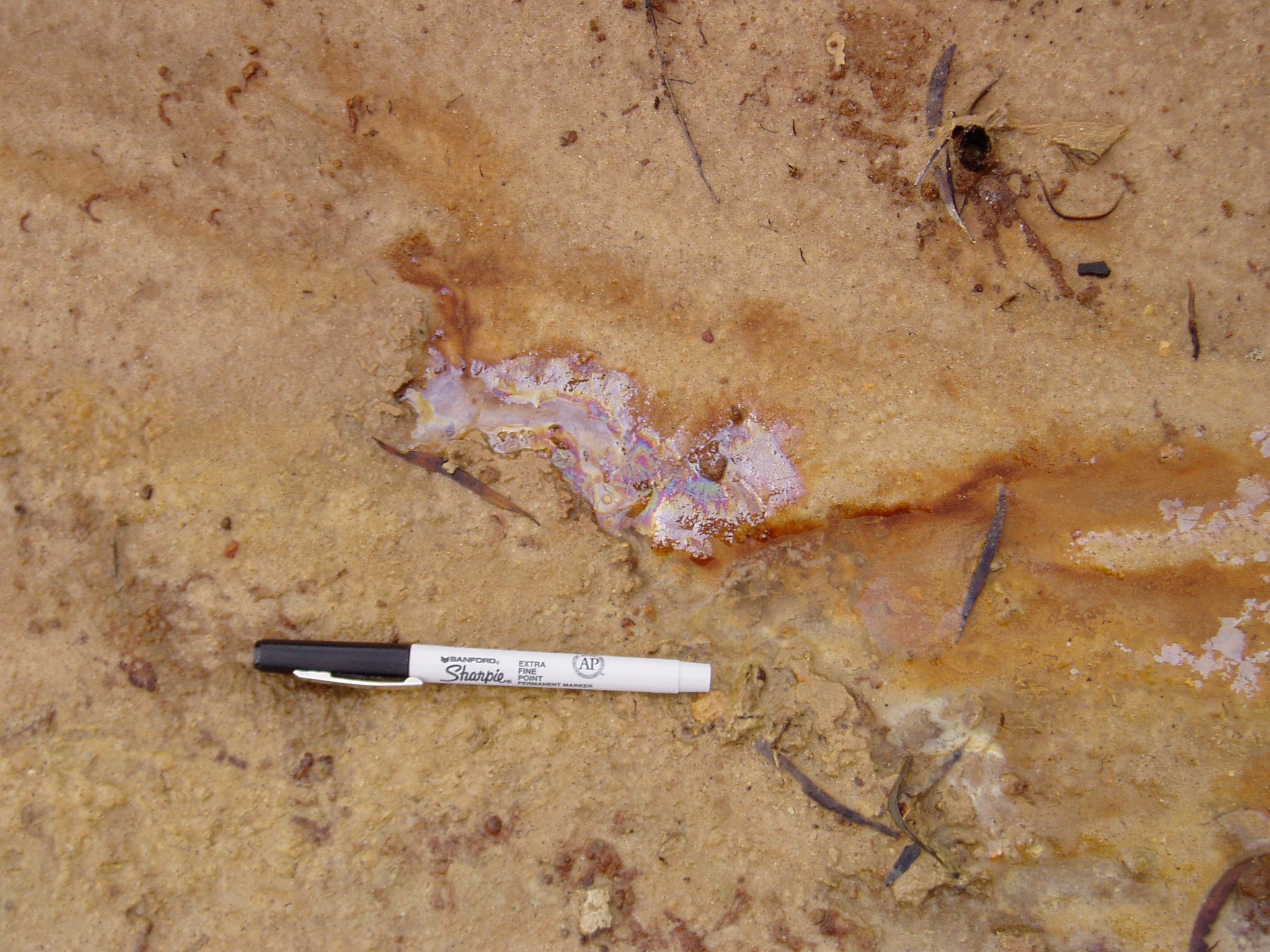
Other indicators of microbial activity include mats, flocculent material, and oily sheen residues. The mats are groups of microbes, usually photosynthetic.
The green color in the left photo indicates that photosynthesis is going on. The middle photo shows white and orange flocculent material. The orange color
is probably iron and the microorganisms are making a rust-like material. The photo on the right shows an oily sheen but it is not oil. Manganese oxidizing
bacteria make this. If you find an oil sheen around a stream, poke it with a stick. If it cracks and breaks, you have found manganese oxide that bacteria
have made!
Photo taken by Bo-young Hong
Photo taken by Melanie Mormile
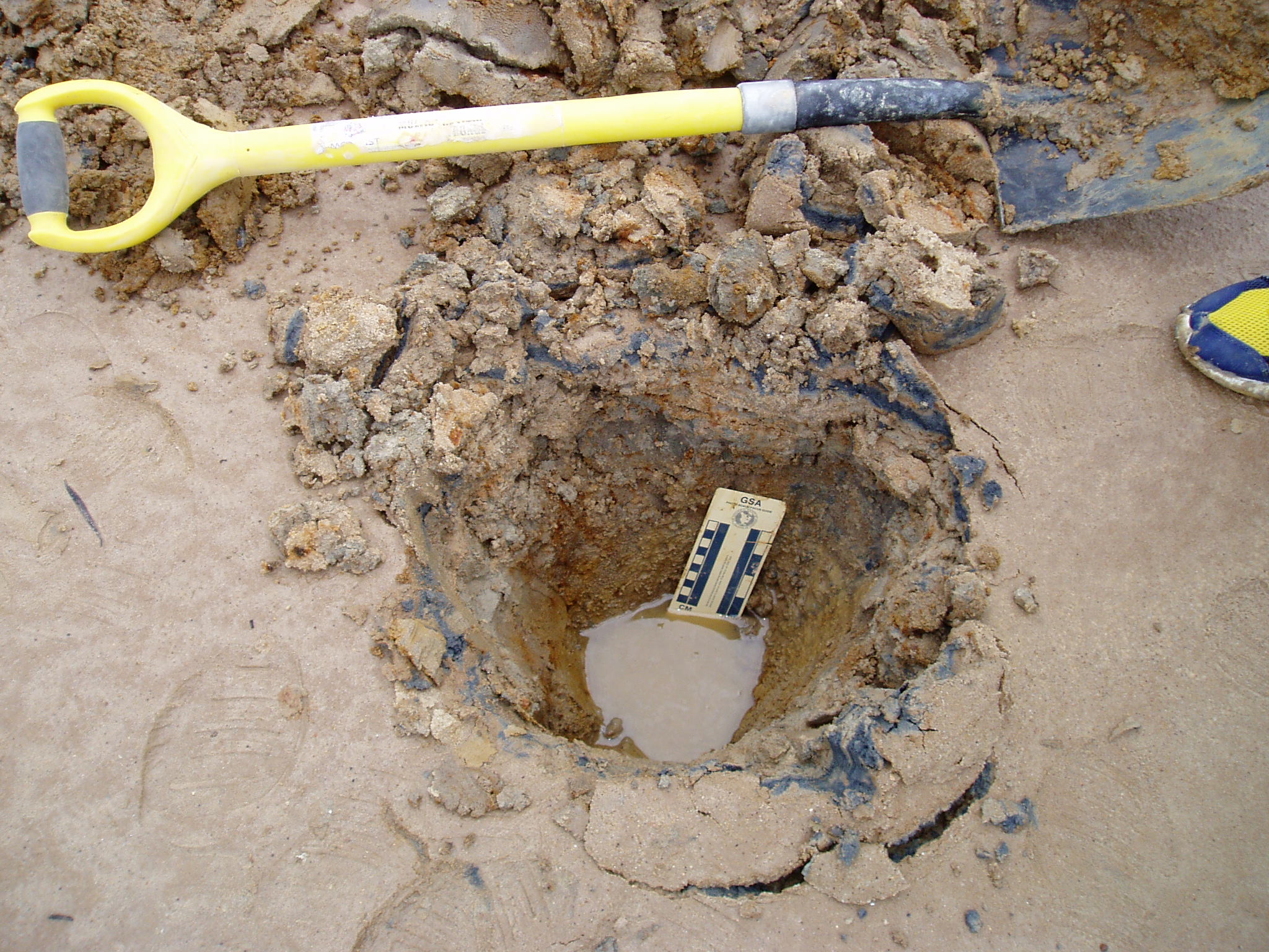
If you dig around a hypersaline lake, you can find other evidence for microbial activity. Some microbes develop different pigments, colors, to help them trap light
for photosynthesis. When you dig up salt crystals, you will usually find that there are layers of colors such as as green, red, purple and black. Each of these colors
represent different microorganisms. Sometimes, when you dig into the mud, you will find black and stinky stuff. This indicates that there are sulfate reducing bacteria
present and they are making hydrogen sulfide, the rotten egg gas.
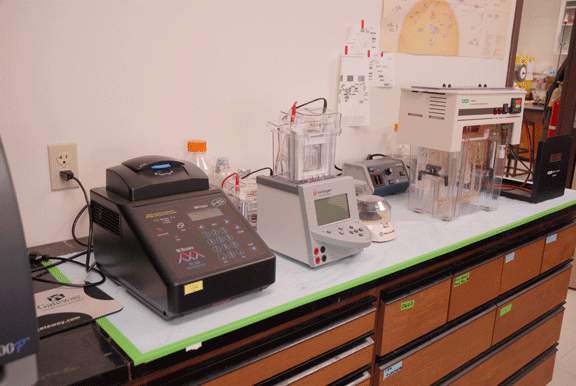
Lab work has to be done though to conclusively show that microorganisms are present in these environments and to identify who they are. This work includes culturing
bacteria and using molecular biology.
Photo taken by Richard Osborn
Some of the equipment that is used to study microorganisms by using their DNA. The machine on the left is a PCR machine. It is used to make many copies of DNA.
To further study the DNA, you need to be able to see it. The machines in the middle and on the right side of the photo are gel electrophoresis machines.
They use electricity to separate out DNA. The resulting gels are stained and UV light is used to see the bands of DNA.
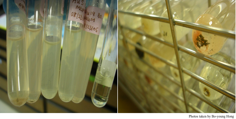
Some of the enrichment cultures that were set-up.
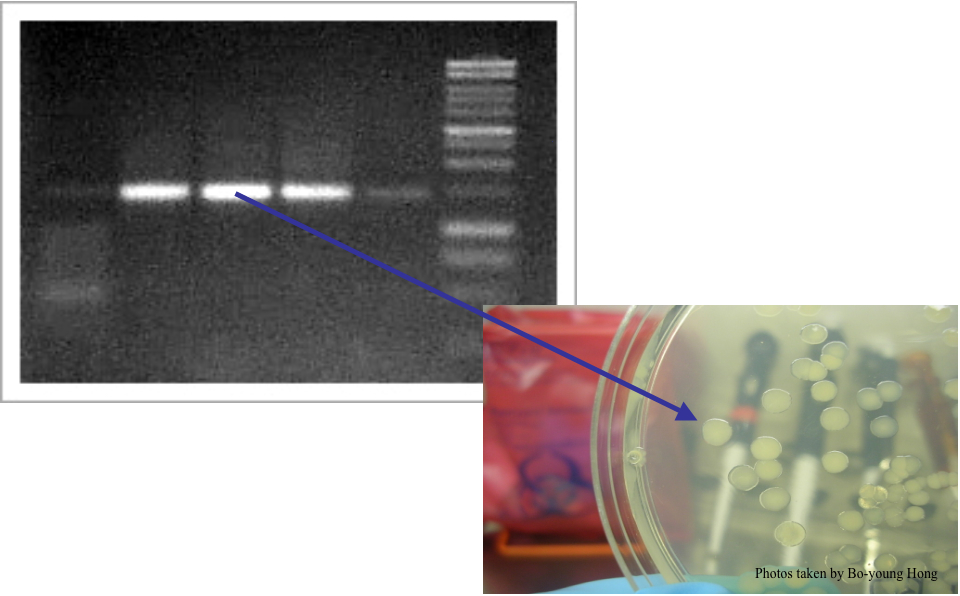
This is a photo of a gel showing the DNA that came from a microbial colony such as the one that is in the bottom photo.